


 http://www.hospitalprocedures.org/sto...
Endotracheal Intubation video
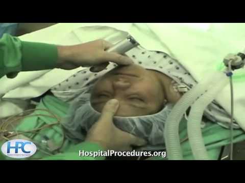
The patient is now being pre-oxygenated with a face mask. A towel has been placed underneath the head just slightly flex the neck. Propofol is being administered as an induction agent. This can slightly burn as it's being administered as is evidenced by the grimacing in the patient's face. She is drifting into a deeper state of anesthesia. You can test for the loss of a lash reflex as a marker for a comatose state, when the patient will need assisted bag valve mask ventilation.
Since the patient is easy to bag valve mask ventilate, a paralytic agent like Succinylcholine can be safely administered. In mask ventilation is then continued until the patient is fully paralyzed. The head is slightly elevated and the neck is extended to place the patient in a sniffing position. A curved Macintosh blade is then inserted into the right side of the mouth, sweeping the tongue to the left, and the tip is placed in the vallecula and the blade is lifted to elevate the epiglottis and expose the subglottic laryngeal structures. You can see the vocal cords in the middle through which the endotracheal tube will be placed and the corniculate tubercles are seen below.
The endotracheal tube is now placed via the right side of the mouth, and should be advanced to a distance of about 21 centimeter at the teeth in women and 23 centimeters at the teeth in men. The endotracheal tubing is now connected to the tube and proper tube placement is confirmed by seeing mist in the tube, symmetric chest wall rise, and carbon dioxide output on an end tidal CO2 monitor. The tube is then secured in place with tape.
Orotracheal Intubation Video procedures and processes | |
| 42 Likes | 42 Dislikes |
| 53,180 views views | 583 followers |
| Education | Upload TimePublished on 1 Sep 2012 |
Không có nhận xét nào:
Đăng nhận xét